 |
Current Projects
Automated Cancer Detection and Grading in Prostate Tissue Histology
A computer aided system for histological prostate cancer diagnosis can assist pathologists by automating two stages. The first is to locate cancer regions in the large digitized tissue biopsy and the second is to assign grades to those regions. In this project we address both of the stages. In the first stage, besides well-known textural features, we use cytological features which are not utilized in the Gleason grading method to detect cancer regions in a whole slide tissue image at 20x magnification (the size is approximately 5,000x20,000 pixels). One of the most important cytological features is the presence of cancer nuclei (nuclei with prominent nucleoli) in the cancer regions. Both cytological and textural features are extracted from the tissue image and are combined to detect cancer regions.
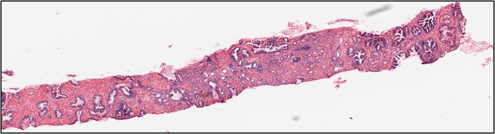 |
| Input whole slide tissue image (5,000x20,000 pixels) at 20x magnification |
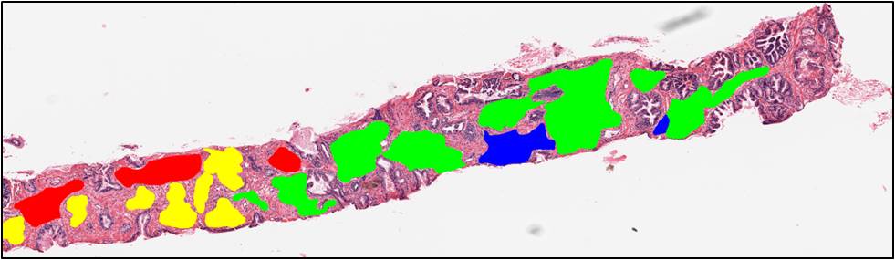 |
| Ground truth of the image. Green regions denote grade 3 cancer, blue regions denote grade 4 cancer. Red and yellow regions denote normal regions. We only consider cancer (blue and green) regions in this project. |
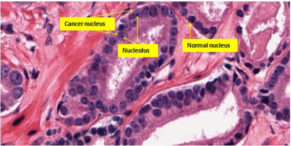 |
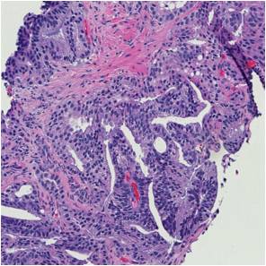
| Cancer nuclei and normal nuclei in the tissue image. A normal nucleus appears with uniform intensity while a cancer nucleus appears light blue with nucleoli (dark spots) inside. |
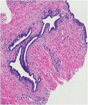
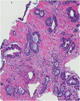
In the second stage, motivated by the Gleason grading method, we aim at classifying a pre-selected region of interest (ROI) into one of the three common categories: benign, grade 3 and grade 4. In the benign ROIs, glands are well-defined with lumina regions in the center, circumscribed by epithelial nuclei and epithelial cytoplasm. In grade 3 ROIs, there appear small, circular glands with thin boundary. Finally, glands start to fuse with each other in grade 4 ROIs. As a result, glands in grade 4 regions do not maintain the well-defined structures as in benign regions. We propose a segmentation-based approach in which we (i) first segment glands from the tissue region, (ii) extract structural features from these segmented glands and (iii) classify the ROI into one of the three classes.
 |
 |
 |
| Benign ROI |
Grade 3 ROI |
Grade 4 ROI |
K. Nguyen, B. Sabata, and A.K. Jain, "Prostate Cancer Grading: Gland Segmentation and Structural Features", Pattern Recognition Letters, Vol. 33, No. 7, pp. 951-961, May 2012.
K. Nguyen, A. Sarkar, and A. K. Jain, "Structure and Context in Prostatic Gland Segmentation and Classification", In: N. Ayache et al. (eds.) MICCAI 2012, LNCS, Vol. 7510, pp. 115-123, Springer, Heidelberg, 2012.
K. Nguyen, A. K. Jain, and B. Sabata, "Prostate Cancer Detection: Fusion of Cytological and Textural Features", Journal of Pathology Informatics, Vol. 2, No. 2 [p. 3], 2011.
K. Nguyen, A. K. Jain, and B. Sabata, "Prostate Cancer Detection: Fusion of Cytological and Textural Features", MICCAI - Workshop on Histopathology Image Analysis, Toronto, Canada, Sep. 18-22, 2011.
K. Nguyen, A.K. Jain and R. Allen, "Automated Gland Segmentation and Classification for Gleason Grading of Prostate Tissue Images", ICPR, Istanbul Turkey, August 23-26, 2010.
|
|
